Surface Computing Support for Image Segmentation
Description
This project is investigating the incorporation of surface computing environments into image segmentation research as an assistive tool using a test driven, software product line approach. Possible directions include:
- Showing that a surface-based assistive tool reduces inter/intra observer variability in image segmentation
- Assessing osteoporosis in pre/post menopausal women, predicting fractures, and developing objective quantitative measures for drug effectiveness for use in pharma trials.
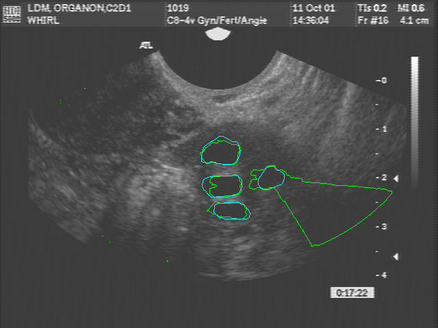
Although the techniques are envisioned to be applicable to a number of areas in health technology, preliminary work is investigating segmentation of ovarian structures. The study of ovarian morphology and function often requires identification, enumeration and measurement of the anatomical structures within the ovary, for example, the follicle outlined in red in the image to the left (follicles are fluid-filled cavities that contain developing eggs). Due to the comparatively low quality of ultrasound images this can be a difficult task. Automated algorithms are sought which can find the precise outlines of these structures while ignoring other structures that may be present in surrounding tissues. These so-called “segmentation” algorithms are crucial for automating analyses for higher-level applications which require knowledge of the number, sizes, and arrangements of ovarian structures. Incorporating surface based computation may support the development of interactive algorithms to reduce inter/intra observer variability.

Partner
- Canadian Light Source


